
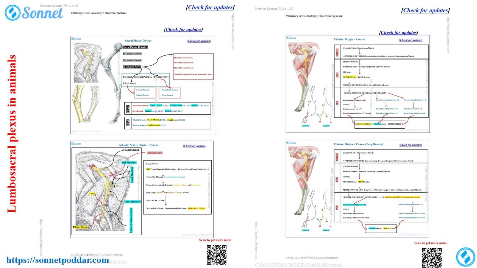
The lumbosacral plexus in animals describes the course and distribution of the nerves from the lumbar and sacral regions. Here, I will provide a summary of the nerves from the lumbosacral plexus of animals, especially of the ox, sheep, goat, horse, and dog.
You will find the concise notes on the lumbosacral plexus nerves of the ox, goat, and sheep. This note might help you to learn the complex courses of the nerves of the lumbosacral plexus easily.
Lumbosacral plexus nerves in animals – ox, sheep, and goat
Here, the lumbar and sacral plexuses are totally different and have specific nerves in these plexuses. First, let’s see the nerves from the lumbar and sacral plexus of the ox, goat, and sheep –
From the lumbar plexus of animals, you will find the following nerves –
- First, the lateral cutaneous femoral nerve
- Second, the femoral nerve, and
- Third, the obturator nerves,
Again, the sacral plexus of the animals presents the following nerves –
- First, the cranial gluteal nerve,
- Second, the caudal gluteal nerve, and
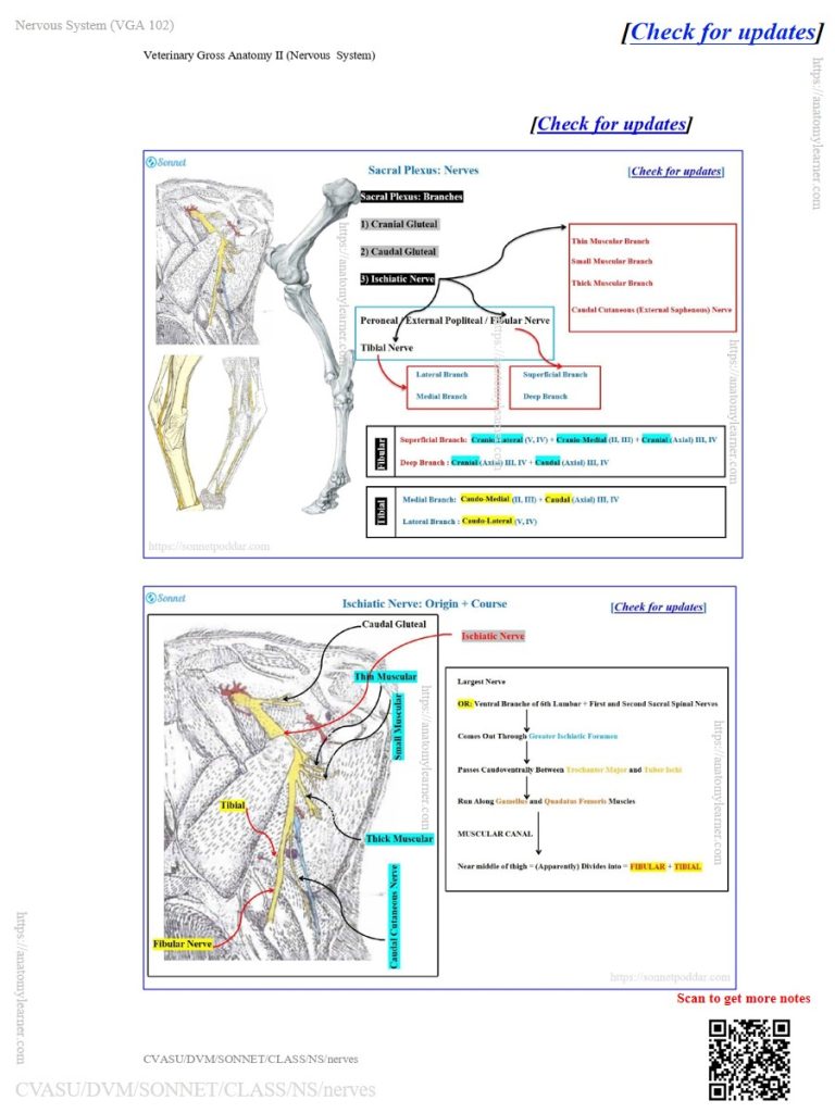
- Third, the ischiatic nerve (the largest nerve of the sacral plexus of the animal’s hindlimb).
However, the ischiatic nerve of the animal’s hind limb has several branches. Let’s identify the branches of the ischiatic nerves from the ox, sheep, and goat’s forelimbs –
- First, a thin muscular nerve,
- Second, a small muscular nerve,
- Third, thick muscular nerve,
- Fourth, the caudal cutaneous sural nerve,
- Fifth, the tibial nerve, and
- Sixth, fibular nerve
NB: Here, I will provide an overview of the notes on the lumbosacral plexus nerves in the animals. Thus, you might go to my other articles and videos to identify the nerves from the lumbosacral plexus of the ox, sheep, and goat.
To identify these nerves, you may follow a good strategy that I have discussed in the notes and diagrams. However, the notes focus on the courses and distribution of the major nerves from the ox, sheep, and goat’s hindlimb with a concise format.
Identification of the lumbar plexus nerve from the ox, sheep, and goat
To identify the nerves from the animal’s lumbar plexus, you might know the followings –
- Bones and segments of the animal’s hindlimb (hip, thigh, leg, and pes regions/segments),
- Important muscles from these 4 segments of the animal’s forelimb,
- Direction and the bones and different terminology related to bones and the hindlimb,
This basic knowledge is also applicable to sacral plexus study.
Here, the first nerve of the animal’s lumbar plexus is the lateral cutaneous femoral/ This nerve arises from the ventral branch of the fourth and fifth lumbar spinal nerves in the ox.
It passes caudal direction between the psoas muscles of the ox. Finally, this nerve extends along the deep face of the subiliac lymph node to reach the skin of the thigh region.
The second nerve of the animal’s lumbar plexus is the femoral nerve, which is the strongest nerve. It arises from the ventral branch of the fifth and sixth lumbar spinal nerves and innervates different muscles of the medial thigh.
This nerve have the clinical important; thus, you need to identify this nerve perfectly.
Finally, the third nerve is the obturator, which innervate obturator externus, gracilis, pectinus, and adductor muscles of the hindlimb.
Nerves of the sacral plexus in animals
The nerves of the animal’s sacral plexus innervate the skin and the caudal lumbar area of the hip, thigh, and leg. I have provided the concise notes on the sacral plexus of the ox, sheep, and goat.
Among the three nerves of the animal’s sacral plexus, the cranial and caudal gluteal are smaller than the ischiatic. However, the cranial and caudal gluteal innervate only the specific muscles.
Cranial gluteal: Innervates the gluteus medius, gluteus profundus, and tensor fascia lata muscles.
Caudal gluteal nerve: Innervates the gluteus profundus, gluteus medius, and gluteus maximus muscles.
However, the ischiatic nerve of the animal’s lumbosacral plexus is the longest and has different branches that innervate through the hindlimb. Again, like the radial, median, and ulnar nerves of the animal’s brachial plexus, you might learn the course of the ischiatic nerve.
I have made the course concise for you to learn the animal’s ischiatic nerve easily. The provided diagrams and notes will help you learn and memorize the course perfectly.
The common questions regarding the course and distribution of the animal’s ischiatic nerves –
- Mention the location of the ischiatic nerve in the hip region of the ox, sheep, and goat.
- Which muscles do you need to remove to get all of the branches of the animal’s ischiatic nerve?
- What are the major branches of the animal’s ischiatic nerve?
- Where does the tibial nerve separate from the fibular nerve?
- Mention the location of the tibial nerve in the leg region of the animals.
- How does the fibular nerve pass from the thigh and leg regions?
- Which digits and surfaces are innervated by the tibia and fibula nerves?
You will be able to answer all the above-mentioned questions if you learn the course of the ischiatic nerve from the provided notes. Here, in the ischiatic nerve course, I have also followed similar learning strategies that I have followed in the brachial plexus nerves.
Ischiatic nerves from the sacral plexus of the ox, sheep, and goat
At first, you should identify the smaller branches from the sacral plexus of the ox, sheep, and goat (identified in the figure). Then, identify the main ischiatic nerve along with the tibia and fibula.
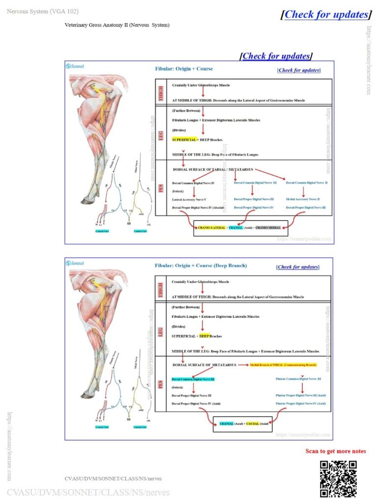
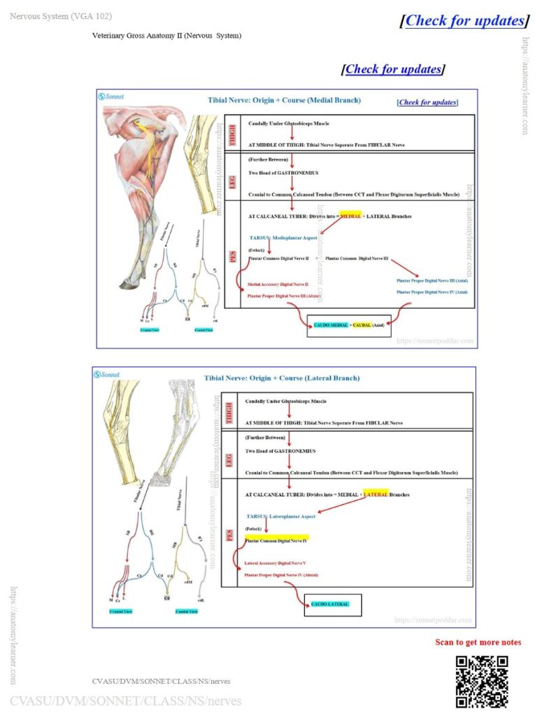
Here, the tibia and fibula remain together and are separated from each other in the middle of the thigh. The tibial nerve lies deep between the two heads of the gastrocnemius muscle of the leg region of the animals.
However, the fibular nerve passes over the lateral head of the gastrocnemius muscle. It deeps along the fibularis longus and extensor digitorum lateralis muscles of the leg region (proximally).
Then, both the nerves (tibia and fibula) present the defined branches and courses in the leg and foot regions. The digits innervate these two nerves from the animal’s lumbosacral plexus.
The innervation of the tibia and fibula to the digits also follows a regular pattern, like radial, median, and ulnar. Here, the diagram presents the surfaces and digits that are innervated by the different branches of the tibia and fibula nerves.
NB: Let’s find the full notes on the animals’ lumbosacral plexus from the veterinary anatomy notes page.
Conclusion
So, the lumbar and sacral plexus form the lumbosacral plexus in animals. Both plexuses provide three major nerves in each.
Here, the iscgiatic nerve is the longest and strongest nerve in the sacral plexus in animals. Its main branches tibia and fibula, innervate the digits of the animal’s hindlimb.
Author