The avian female reproductive system consists of the ovary and the oviduct. Here, I will describe all the features of the avian ovary and different parts of the oviduct with a diagram.
However, I will provide a concise note and diagram on the anatomy of the organs from the avian female reproductive system.
Special features of the avian female reproductive system
Let’s see the special features of the avian female reproductive organs –
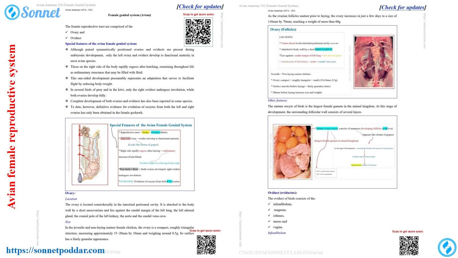
- During embryonic development, both the ovaries and oviducts (right and left) developed symmetrically. However, only the left ovary and oviduct remain functional throughout life.
- The right oviduct of the avian species rapidly regresses and remains as a rudimentary part throughout life. However, this rudimentary part of the oviduct remains filled with the fluid.
- This one-side oviduct and ovary have an indirect effect on flight. This is due to the reduction of the body weight of the avian species.
- In some of the avian species, the right oviduct undergoes involution. However, both ovaries develop fully in these species (prey bird and kiwi).
- Again, full development of both ovaries and oviduct may also be found in some of the avian species.
For example, Goshawks have fully developed ovaries on both the right and left sides. However, the ovulation of the oocyte occurs from both the right and left ovaries.
Now, let’s describe the anatomical features of the ovary and oviduct from the avian species separately.
Avian ovary anatomy (chicken, turkey, duck)
Here, I will discuss the important features of the avian ovary anatomy with a diagram. But you might have the basic idea of the mammal’s ovary anatomy (for example: cow ovary, sow ovary, ewe ovary, queen ovary and others).
Let’s see the unique anatomical features of the avian ovary –
Location of the avian ovary
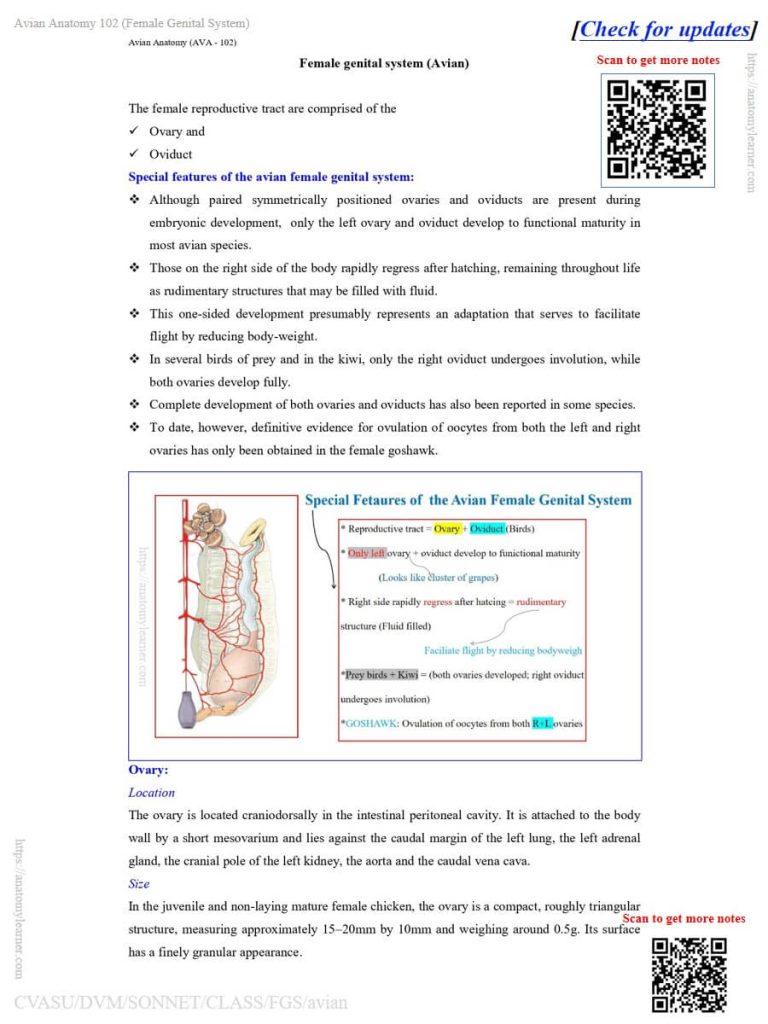
The avian ovary, for example chicken ovary, is located craniodorsally in the intestinal peritoneal cavity. It is attached to the chicken’s body wall by the short mesoovarium.
However, the chicken ovary lies against the caudal margin of the left lung, the left adrenal gland, the cranial pole of the left kidney, the aorta, and the caudal vena cava.
Size of the avian ovary – chicken ovary
The avian ovary is variable in different avian species. However, the size is also variable between the juvenile and non-laying birds.
It is compact and roughly triangular in the juvenile and non-laying mature chicken. They are 10×20 millimeters in length and weigh around 0.5 grams. You will find the fine granules’ appearance on the surface of the avian ovary.
As the ovarian follicle matures after laying, the ovary increases its size and weight. You will typically get 110×70 millimeters in length and a weight of about 60 grams of an avian ovary.
Other features of the avian ovary/chicken ovary
A mature oocyte of an avian species is the largest female gamete in the animal kingdom. At the developmental stage, you will find several layers in the surrounding follicular wall of the ovary.
Here, the provided notes and diagrams of the avian female reproductive system might help you to learn these features perfectly. However, the note provide details description of the anatomy of the avian ovary and also the avian oviducts with the proper diagrams.
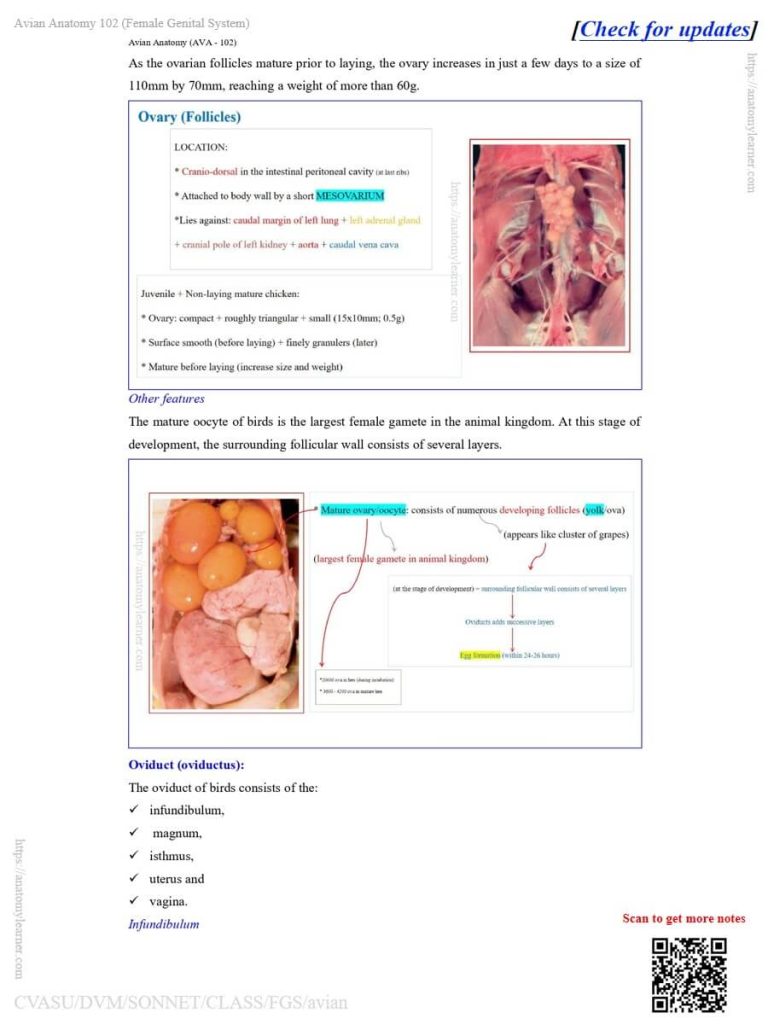
Parts of the oviducts in birds or avian species
First, let’s see the diagram of the parts of the oviducts in birds or avian species. This is very important as you need to learn and draw every single part of the avian oviduct.
Here, the avian oviducts have the following different parts –
- Infundibulum part,
- Magnum part,
- Isthmus part,
- Uterus part, and
- The last part (shown on the diagram).
Now, I will describe all the parts of the avian oviducts with the diagrams.
Questions on the avian female reproductive organs
However, you should know the question pattern of the avian female reproductive system. Let’s see the possible questions for the theory examination –
- What are the special features of the avian female reproductive organs?
- Mention the location, size, and unique features of the chicken ovary with a diagram.
- Draw and label the various parts of the avian/chicken oviduct.
- Describe the egg formation process in the chicken.
- How do the various parts of the chicken oviduct contribute to form a egg?
Thus, while learning the anatomical features of the different parts of the chicken oviduct, you might also learn their function. Thus, you may easily answer the last two questions that I provided in the above list.
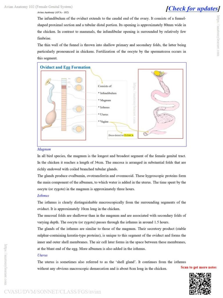
Infundibulum of the chicken oviduct
Here, the infundibulum is the first part of the chicken oviduct and differs from that of mammals. It extends to the caudal end of the ovary and attaches to the magnum.
The infundibulum of a chicken has a funnel-shaped proximal section and a distal tubular part. Here, the infundibulum possesses relatively fewer fimbriae.
However, the wall of the funnel is thin and possesses the primary and secondary folds.
Contribution to egg formation: Fertilization of the oocyte by the spermatozoa occurs in this segment.
Magnum of the chicken oviduct
It is the second segment and the continuation of the chicken oviduct caudally. It is the longest and broadest segment of the avian female reproductive tract. However, you will find approximately 35 centimeters in the magnum of a chicken.
Here, the mucosa of the avain magnum possesses substantial folds which consist of coiled branched tubular glands. However, these glands produce ovalbumin, ovotransferrin and ovomucoid.
During egg formation, the time spent by the oocyte in the magnum is approximately three hours.
Isthmus of the chicken oviduct
Grossly, the isthmus is part of the connection between the magnum and uterus segment of the chicken oviduct. However, you can also differentiate the isthmus of the chicken oviduct from the magnum by their internal mucosal folds.
Here, the mucosal folds of the chicken’s isthmus are shallower than the magnum. They are associated with the secondary folds of varying depth.
Contribution of the isthmus segment: Here, the oocyte passes through the isthmus in around 1.5 hours. You will find a similar type of gland in the isthmus, like those of the magnum.
These glands secrete the stable sulphur-containing keratin-type proteins. However, it is unique to this segment of the oviduct. This type of secretion helps to form the inner and outer shell membranes of the hen’s egg.
There is an air cell at the blunt end of the egg, which forms later in the space between the outer and inner shell membranes. However, within the isthmus, more albumen is also added.
Uterus of the avian oviduct
The uterus of the avian oviduct is another important segment, which is also termed the shell gland. It is the continuation of the isthmus caudally and about 8 centimeters long in an ideal chicken.
The uterus in the avian, or chicken, expands into a pouch-like structure. Here, the muscular layer is well-developed in the chicken oviduct.
Contribution of the uterus: In this segment of the avian oviduct, eggs spend more time (around 20 hours). Thus, the time spent is longer in the uterus than in any other segment of the avian oviduct.
It forms the calcareous shell from calcium carbonate and other calcium salts. However, the cuticle of the egg is also derived from the uterus of the chicken or the avian oviduct. Here, the cuticle is the thin, organic outermost layer of the egg.
How is the egg formed within the chicken oviduct?
To describe the egg formation within the chicken oviduct, you might focus on two points –
- Yolk = comes from the ovary, and
- Albumen, shell membrane, and egg shell = come from the various parts of the chicken oviduct.
Here, the provided diagram and note describe the details process of egg formation. After ovulation yolk drops into the oviduct, where the magnum of the oviduct secretes white albumen around the yolk.
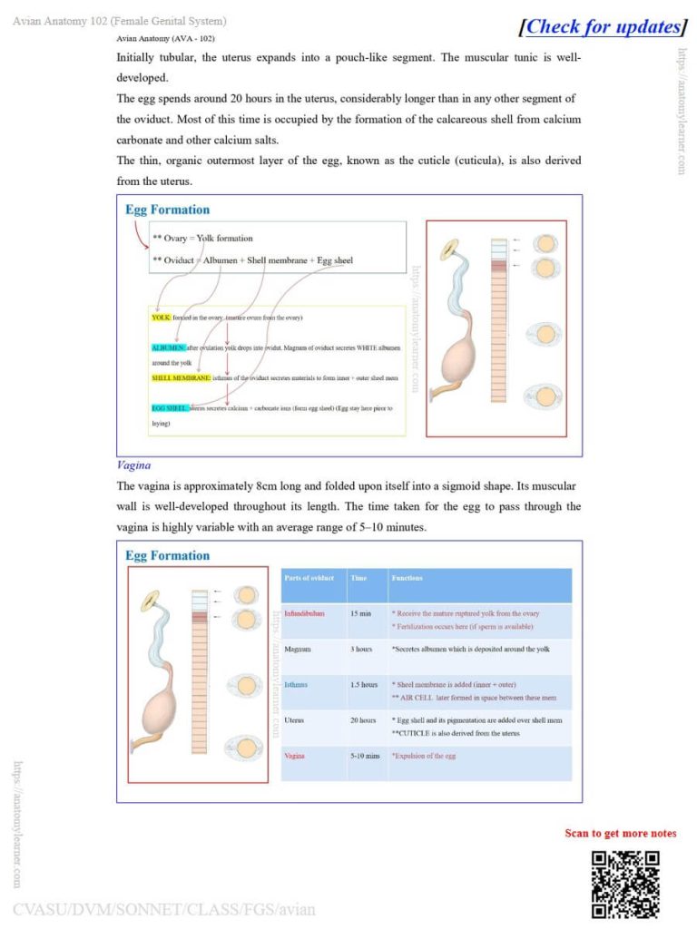
The isthmus of the oviduct secretes the materials to form the inner and outer shell membranes. However, the uterus secretes the calcium and carbonates, which form the egg shell.
You might also mention the time spent in various segments of the chicken oviduct during the formation of an egg.
FAQ’s on the hen reproductive system
Well, it requires 24 hours to form an egg in a hen. However, during the formation of an egg, it spends 15 minutes, 3 hours, 1.5 hours, 20 hours, and 10 minutes in the infundibulum, magnum, isthmus, uterus, and last segment, respectively.
When the oocyte passes through the isthmus of the hen’s oviduct, it forms the air cell at the blunt end of the egg. However, this air cell is formed between the inner and outer shell membranes of the hen’s egg.
Conclusion
So, the avian female reproductive system is unique than these of the mammals and consists of only an ovary and oviduct. Here, the ovary or ovarian follicles are also unique in the avian species.
However, the oviduct consists of 5 different segments which are also unique compared to those of the mammal’s uterus.
References
- Rutllant and Khamas, (2024). Reproductive System – Anatomy and Histology of the Domestic Chicken.
- Mothiba LM, Sathekge LJ, Hlokoe VR, and Tyasi TL (2025). Morphometry of the Reproductive System in Boschveld Chickens. Worl. Vet. J., 15(3): 703-709. DOI: https://dx.doi.org/10.54203/scil.2025.wvj71
- Rahman, M. A. (2014). An Introduction to the Morphology of the Reproductive System and the Anatomy of the Hen’s Egg. Jr of Life and Eart Sci., 8, 1–10.
Written by