This article will help you identify the surface epithelium of animals under the microscope. Before you start, ensure you are comfortable using a light microscope in a histology laboratory.
If you need a foundation in permanent slide preparation, refer to my previous article. This guide focuses on helping beginners recognize the different types of surface epithelium. For tips or a review on microscope use, consult my other article.
“This article is not for the expert. It’s only for the newbie.”
Requirements
You should have a basic idea of acidic and basic dyes and their activities on animal tissue. You should also be familiar with permanent slide preparation and the use of a light microscope.
- You should know the definition of tissue, the four basic types of tissue, and the characteristics of epithelium tissue.
- You should know the basement membrane.
- Finally, you should know different types of surface epithelium (classification of surface epithelium)
Classification of surface epithelium
Lining epithelium is divided into two major types – surface epithelium and glandular epithelium. In this article, our focus is on identifying the surface epithelium of the animal body, which covers all external and internal surfaces. With this distinction in mind, we will proceed to identify various types of surface epithelium under the microscope.
Simple epithelium (presence of a single layer of epithelium cells on the basement membrane)
- Simple squamous epithelium
- Simple cuboidal epithelium
- Simple columnar epithelium
Stratified epithelium (presence of two or more layers of epithelium cells on the basement membrane)
- Stratified squamous epithelium
- Stratified cuboidal epithelium
- Stratified columnar epithelium
- Transitional epithelium
Pseudo-stratified epithelium has a single layer of cells on the basement membrane, but not all cells reach the apical surface. Nuclei are present at different levels.
Pseudo-stratified columnar epithelium
“This classification is based on the number of layers and the shape of epithelium cells.”
Let’s identify and analyze each type of animal surface epithelium under the microscope.
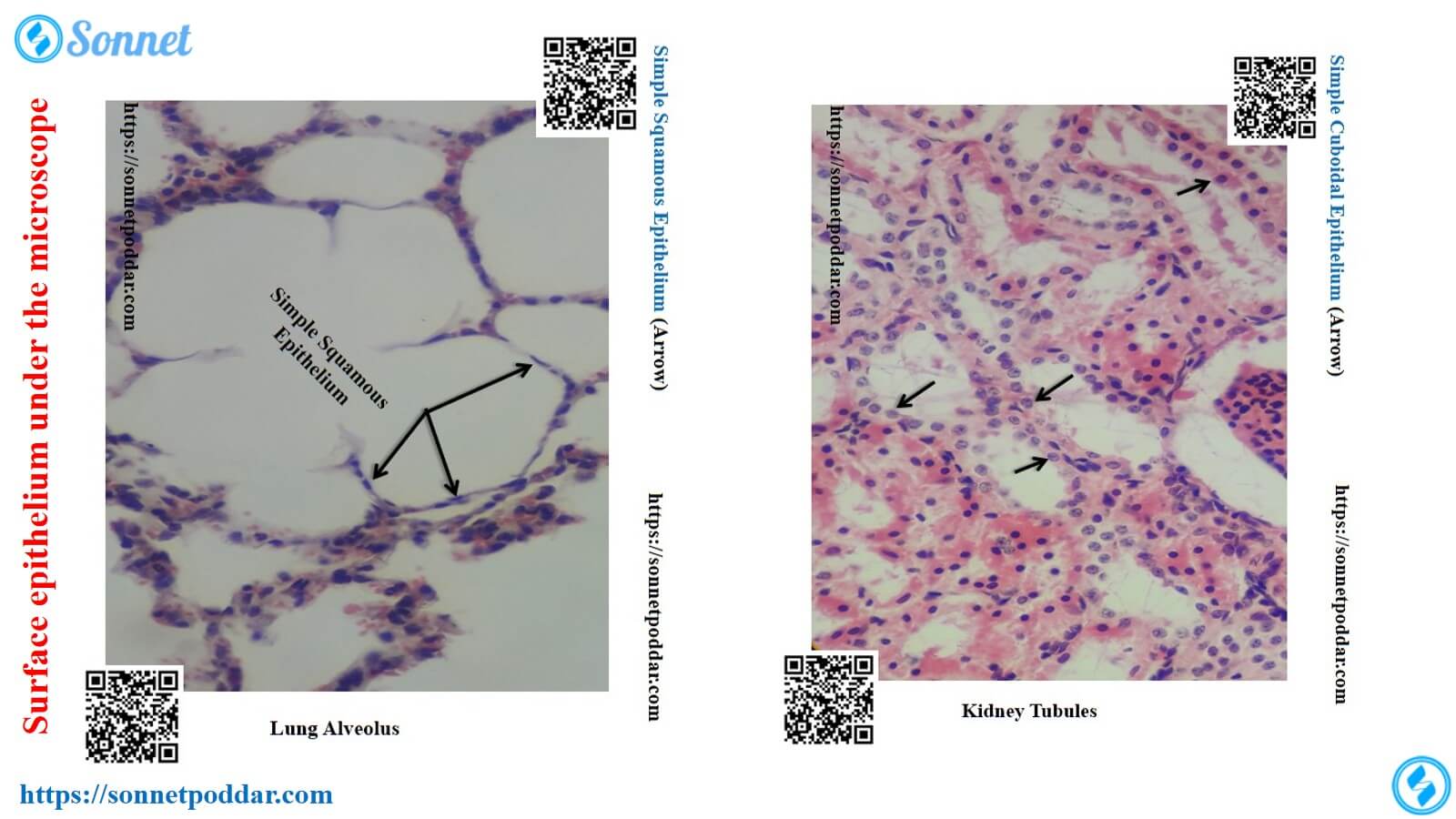
Simple squamous epithelium
We will find the following characteristics of simple squamous epithelium under the microscope –
- Presence of a single layer of cells
- The cells are thin, flat, plate-like, and shaped like polygons.
- There is a flat nucleus placed in the center.
- In the surface view of simple squamous epithelium, cells look polygonal with serrated borders. The nucleus is oval or spherical and present in the cell center.
Location of simple squamous epithelium
You will find simple squamous epithelium on –
Lining of the serous membrane of the body cavity (pleural cavity, pericardial cavity, and peritoneal cavity – they are also called mesothelium)
Lining of the heart, blood vessels, and lymphatic vessels (also referred to as endothelium)
Simple cuboidal epithelium
We will find the following characteristics of simple cuboidal epithelium under the microscope –
- Presence of a single layer of cells
- The height and width of the cell are almost equal.
- Presence of a centrally placed, rounded nucleus
There are also variations: low cuboidal epithelium (cells shorter than wide) and tall cuboidal epithelium (cells taller than wide).
Location of simple cuboidal epithelium
You will find simple cuboidal epithelium on –
- Lining the follicle of the thyroid gland
- Lining the ducts of many glands
- Lining the collecting duct of the kidney
- Lining the germinal epithelium of the ovary
Simple columnar epithelium (Ciliated or non-ciliated)
We will find the following characteristics of simple columnar epithelium under the microscope –
- Presence of a single layer of cells
- The cells are much taller than they are wide and are narrow.
- The apical surface of these cells may have cilia. With cilia, the cells form ciliated simple columnar epithelium. Without them, they are non-ciliated columnar epithelium.
- The nucleus is oval or long and sits near the basement membrane.
Location of simple columnar epithelium
You will find simple columnar epithelium on –
- Lining of the glandular stomach, intestine of an animal
- The lining of the uterus and the uterine tube of an animal
Stratified squamous epithelium (keratinized or non-keratinized)
There are two types of stratified squamous epithelium: keratinized and non-keratinized.
We will find the following characteristics of stratified squamous epithelium under the microscope –
- Presence of several layers of cells
- The top layer of cells looks like simple squamous epithelium. They are thin and flat with flat nuclei.
- The cells in the deep layers are polygonal in shape.
- If keratinized, non-nucleated flat cells fill with keratin. This forms a hard protective barrier.
Location of stratified squamous epithelium
You will find stratified squamous epithelium on the following organs –
- Keratinized stratified squamous epithelium – skin of the animal body
- Non-keratinized squamous epithelium – esophagus, tongue, vagina
Stratified cuboidal epithelium
We will find the following characteristics of stratified cuboidal epithelium under the microscope –
- Presence of two or more layers of cells
- Cells show characteristics of simple cuboidal epithelium. Height and width are almost equal. Nuclei are round and centrally placed.
Location of stratified cuboidal epithelium
You will find stratified cuboidal epithelium on the following organs of the animal’s body-
- The lining of the sweat gland of an animal (Skin)
- Lining of the salivary gland
The name of stratified epithelium is based on the shape of the superficial cell layers. It is not based on the shape of deeper layers.
Stratified columnar epithelium
We will find the following characteristics of stratified columnar epithelium under the microscope –
- Presence of several layers of cells
- The superficial cells are taller than they are wide and are narrow. They look like simple columnar epithelium.
- Deeper cells are small, polygonal, or columnar. They do not reach the surface.
Location of stratified columnar epithelium
You will find stratified columnar epithelium on the following organs of the animal’s body –
- Lining epithelium of the distal portion of the urethra
- Lining of the duct of the mandibular gland
- Lining epithelium of the duct of the parotid gland
Pseudo-stratified columnar epithelium
As noted, pseudo-stratified epithelium consists of a single layer on the basement membrane. Nuclei at different levels create a pseudo-stratified appearance.
You will find the following characteristics of pseudo-stratified columnar epithelium under the microscope –
- Presence of single-layer, irregular shaped cell
- Presence of elongated or oval nuclei, and they are located at different levels
- The top surface of these cells may have cilia or stereocilia.
- Presence of a goblet cell
Location of pseudo-stratified columnar epithelium
You will find pseudo-stratified columnar epithelium on the following organs –
- Ciliated pseudo-stratified columnar epithelium – lining of trachea, larger bronchi
- Lining epithelium of epididymis and ductus deference (presence of stereocilia; does not contain goblet cells)
Transitional epithelium
You will find the following characteristics of transitional epithelium under the microscope –
- Presence of several layers of cells
- The superficial cell layer changes with the state of the organ. In a relaxed state, pillow-shaped cells have round nuclei. When stretched, these cells become flat and elongated with flattened nuclei.
- Presence of smaller and irregular shaped cells at deeper layers
Location of transitional epithelium
You will find transitional epithelium on the following organs –
- Lining of the urinary bladder
- Lining of the ureter
- Lining of the urethra
Conclusion
You now have a clear framework for identifying the surface epithelium of an animal under the microscope. But you might know the details of different types of surface epithelium of animals.