
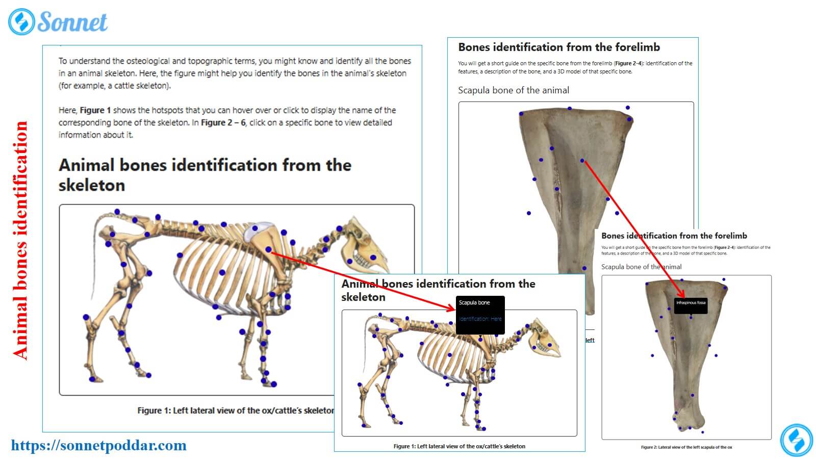
To understand the osteological and topographic terms, you might know and identify all the bones in an animal skeleton. Here, the figure might help you identify the bones in the animal’s skeleton (for example, a cattle skeleton).
Here, Figure 1 shows the hotspots that you can hover over or click to display the name of the corresponding bone of the skeleton. In Figure 02 – 60, click on a specific bone to view detailed information about it.
Animal bones identification from the skeleton
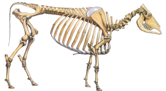

Figure 1: Left lateral view of the ox/cattle’s skeleton
N. B. The materials (real samples) provided here are intended solely for practicing the osteological features of the animal’s bones. Thus, you are expected to have the prior knowledge acquired from both theoretical and practical classes.
If you are accessing these materials from your mobile, please switch to desktop site mode. This is to ensure that the hotspots on the bones are displayed and viewable.
When you hover over the hotspot of every main bone in Figure 1, a pop-up box shows some of the linked contents. However, Figure 02-60 shows hotspots exhibiting various osteological features from the major bones of the skeleton.
Bones identification from the forelimb
You will get a short guide on the specific bone from the forelimb (Figure 02-15): identification of the features, a description of the bone, and a 3D model of that specific bone.
Scapula bone of the animal
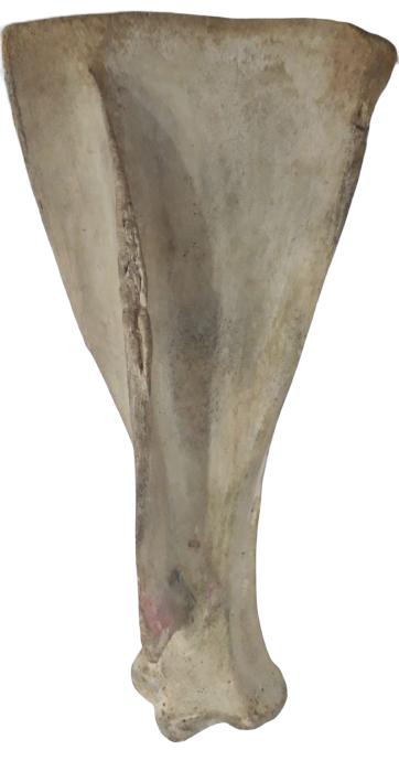
Figure 2: Lateral view of the left scapula of the ox
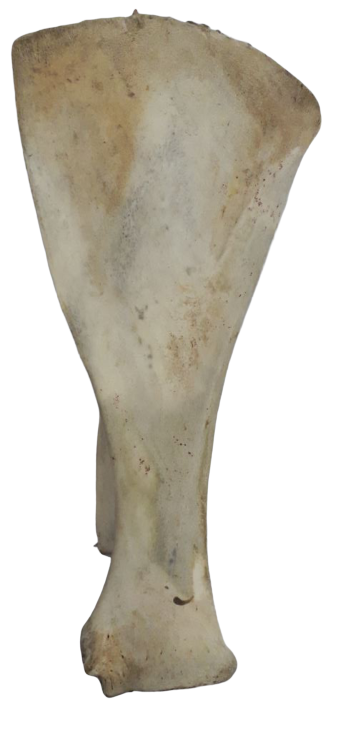
Figure 3: Medial view of the right scapula of the ox
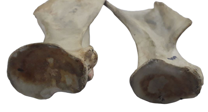
Figure 4: Distal extremity of the scapula (Left and Right scapula bones + Medial and lateral views)
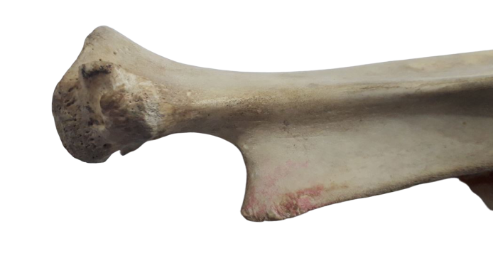
Figure 5: Distal extremity of the left scapula (Medial and lateral views)
Humerus bone of the animals (Ox, Horse, Dog)
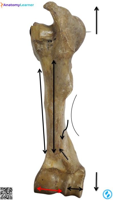
Figure 6: Left humerus of the animals (Ox) – Anterior view
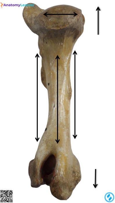
Figure 7: Left humerus of the animals (Ox) – Posterior view
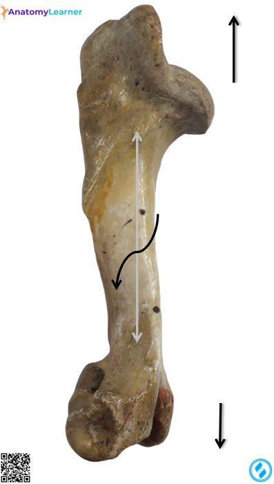
Figure 7: Left humerus of the animals (Ox/horse) – Lateral view
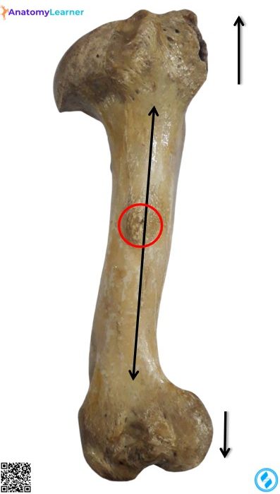
Figure 8: Humerus of the ox (Medial view)
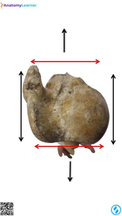
Figure 9: Left humerus of the animal/ox/horse (Proximal extremity)
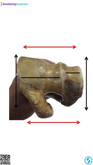
Figure 10: Left humerus of the animal/ox/horse (Distal extremity)
Animal radius bone (Ox, horse, pig, and dog)
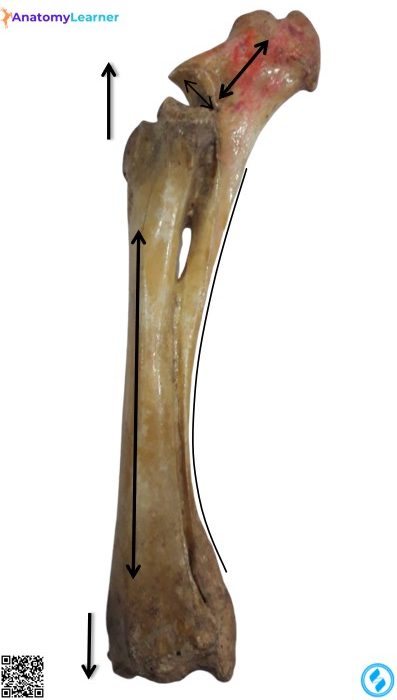
Figure 11: Animal radius bone (Anterolateral view)
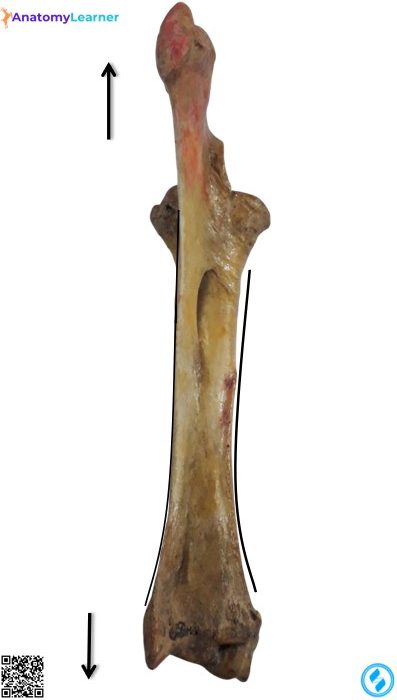
Figure 12: Animal radius bone (Posterior view)
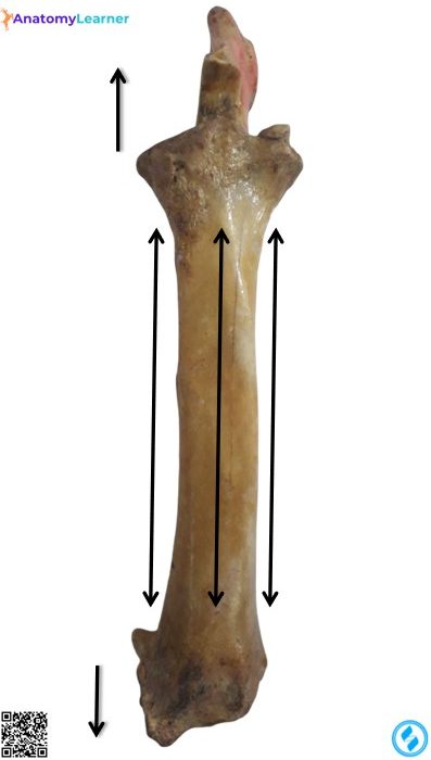
Figure 13: Ox radius bone (Anterior view)
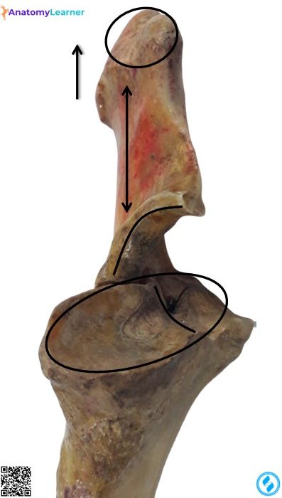
Figure 14: Proximal extremity of the animal radius and ulna bones
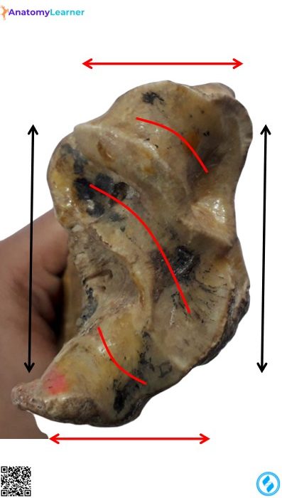
Figure 15: Distal extremity of the animal’s radius and ulna bones
Metacarpal bones of the animals
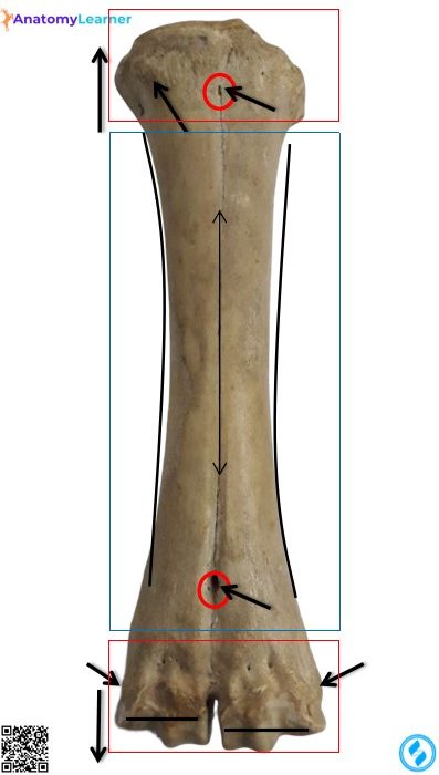
Figure : Dorsal view of the animals metacarpals (Ox, horse, pig, and dog)
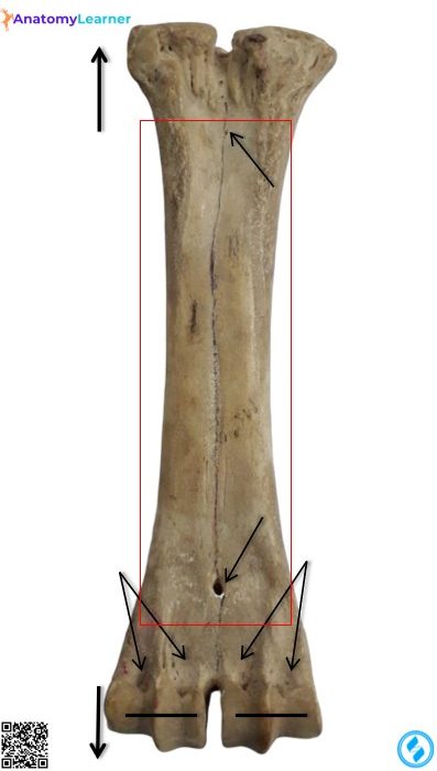
Figure: Palmar view of the animals metacarpals III and IV (Ox, horse, dog)
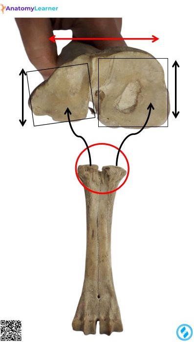
Figure: Proximal extremity of the animals metacarpals (Ox, horse, dog)
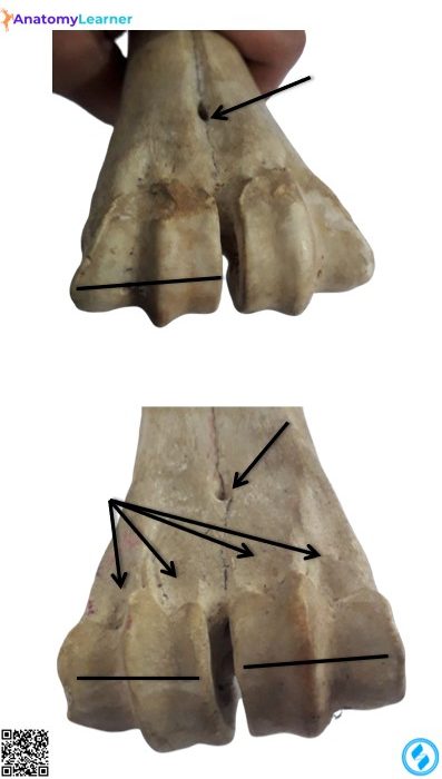
Figure: Distal extremity of the metacarpals of the animals (Ox, horse, dog)
Bones identification from the animal’s hindlimb (Ox, horse, and dog)
Femur bone of the animals
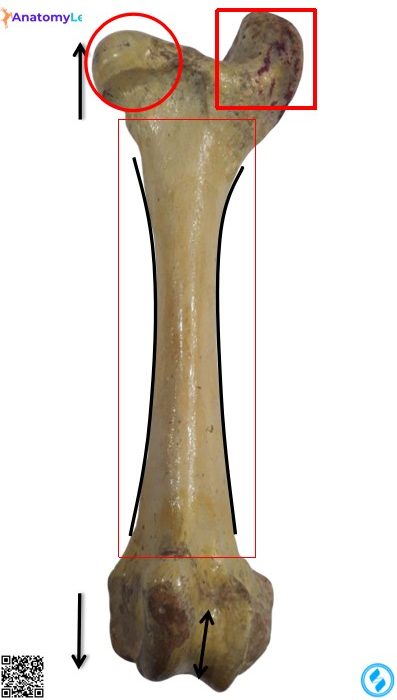
Figure: Anterior view of the animal’s femur bone (Ox, horse, and dog)
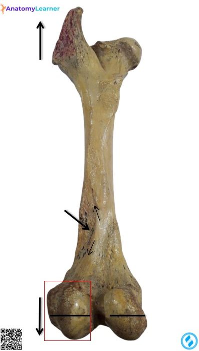
Figure: Posterior view of the animal’s femur bone
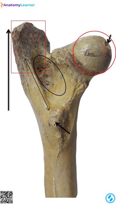
Figure: Proximal extremity of the animal’s femur bone (Ox, horse, dog)
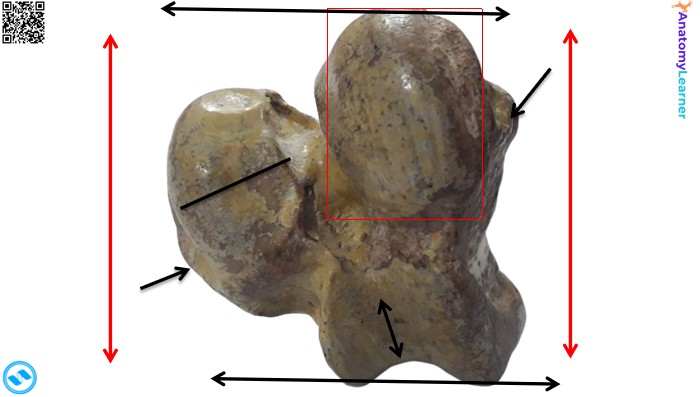
Figure: Distal extremity of the femur bone (Ox, horse, dog)
Tibia and fibula bones of the animal
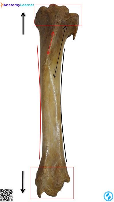
Figure: Anterior view of the tibia bone (Ox, horse, dog)
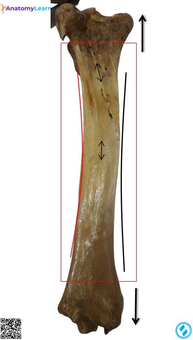
Figure: Posterior view of the animal’s tibia bone (Ox, horse, dog)
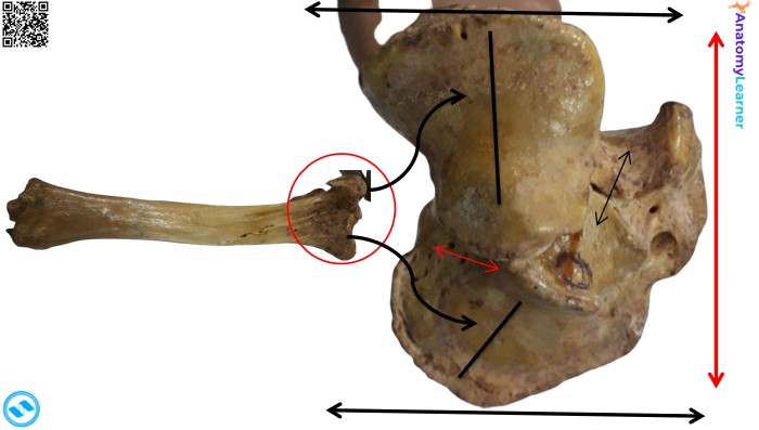
Figure: Proximal extremity of the animal’s tibia bone
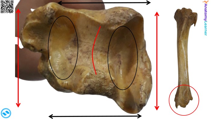
Figure: Distal extremity of the tibia bone (Ox, horse, dog)
Animal metatarsal bones
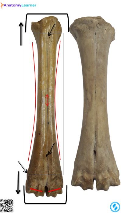
Figure: Dorsal view of the metatarsal bones (Ox, horse, dog)
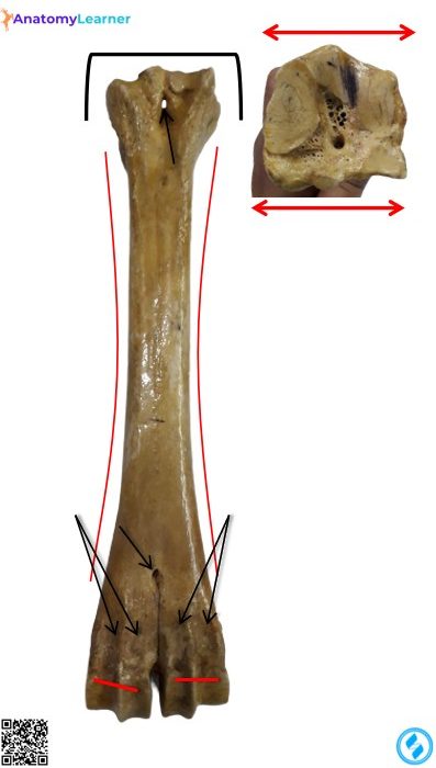
Figure: Planter view of the metatarsal bones (Ox, horse, dog)
Animal vertebrae (Ox, horse, pig, and dog) identification
Typical vertebra of the ox, horse, pig, and dog
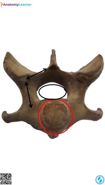
Figure 16: Typical vertebra of the animal (Cranial view)
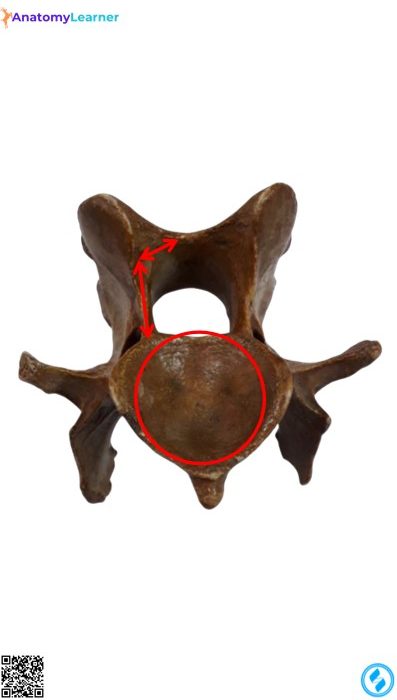
Figure 17: Animal’s typical vertebra (Caudal view)
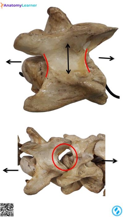
Figure 18: Ox vertebrae (Lateral views)
Animal atlas vertebra bone
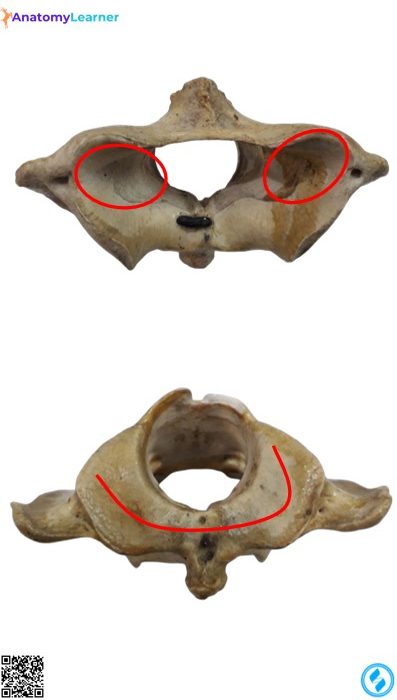
Figure 19: Atlas/ First cervical vertebra of ox, horse, pig, and dog (Anterior and posterior views)
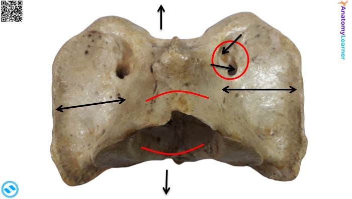
Figure 20: Dorsal view of the animal’s atlas vertebra bone
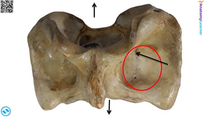
Figure 21: Ventral view of the animal’s atlas vertebra bone
Animal’s second cervical vertebra (Axis bone)
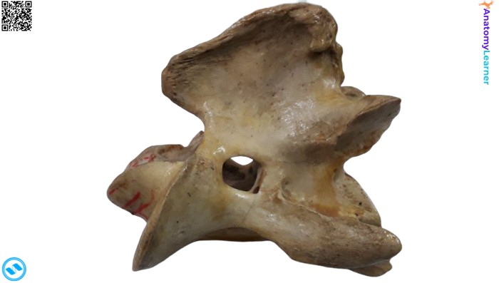
Figure 22: Animal’s second cervical vertebra (Lateral view)